5 months ago I woke up with the anxiety, fear and hope that
I was beginning the ending of almost a year’s worth pain and extremely limited
mobility/quality of life.
I was wheeled in to an operating room and 7 hours later I
woke to what I was told was a very successful surgery. My labrum had been
detached which was more severe than expected the repair went well. I was told I
had good joint space and that the reshaping of my femoral head & neck was
smooth. I was also told that my acetabulum was now structurally sound. The
periactetabular osteotomy or PAO looked great. I had seven screws holding my
“new” hip together as I re-learned how to move it. Initially, things appeared
to be going well and the more detailed history can be read below. There were
some complications that seemed mild and some typical trip-ups along the way of
recovery which was starting to seem slower than some of my new “hippie”
friends. I asked questions and I followed my “rules.” After 8 weeks (the
typical initial recovery) I went in for a follow-up appt. A set of x-rays
determined that my cuts appeared to be healing but “not quite there yet.” And I
was told I needed another 2 weeks on crutches. I was cleared after that and
started physical therapy.
Which brings me where I am today. Thought the next 2.5
months of PT and beginning to bear weight I continued to have a significant
amount of pain. Which after pushing my
me and therapist resulted in the last two blog entries discussing some more
imaging results of a MR-Arthrogram (MRI with contrast) and a CT.
As I was driving home two weeks ago, Dr. Prevost called me
to give the results of the CT scan. He said that after reading the
radiologist’s report and thoroughly reviewing the images himself he agreed with
that fact one of the breaks in my pelvis was not healing. This is considered
delayed or non-union. He used the term non-union as he talked with me and went
on to explain that it is very rare (occurs in about 1% of PAO surgeries), he
has never physically seen it and he didn’t know what to do about it (if
anything). He wanted to send the images down to Dr. Mayo immediately and talk
with him regarding next steps. “The next
decisions will be Dr. Mayo’s,” he said. He also said that it would cause
significant pain but he still wasn’t sure what they would do (bone graft, plate
& screws), if anything because often they won’t do anything for about a
year to be sure the bones are not healing.
There has been several brief conversations emailed back and
forth with Kenda, Dr. Mayo’s PA-C and as far as I know, he has the images and
is going to be contacting me. I don’t know when.
I am having a hard time dealing this emotionally right now. Other
things have been mentioned as possibilities for my pain like, issues with the
labral repair or a new tear, the PSOA tendon and just plain old slow recovery.
I KNOW something is not right. This pain started from the labral tear – I was
told that repairing the labrum would not fix the problem because of the
structure of my bones. I was told that they wouldn’t even consider fixing just
the labrum and I need the PAO. I went for it, not expecting for a miracle but
hoping that in 3-6 months post-op I would be A LOT better than I was at that
time. Well I am not. I feel as though I have recovered surgically from one of
the most invasive orthopedic surgeries out there but I am still in an amount of
daily CONSTANT pain that is comparable (and only slightly different) then
pre-op and now the left side is hurting. Leftie was worse off according to
imaging a year ago and that scares me too.
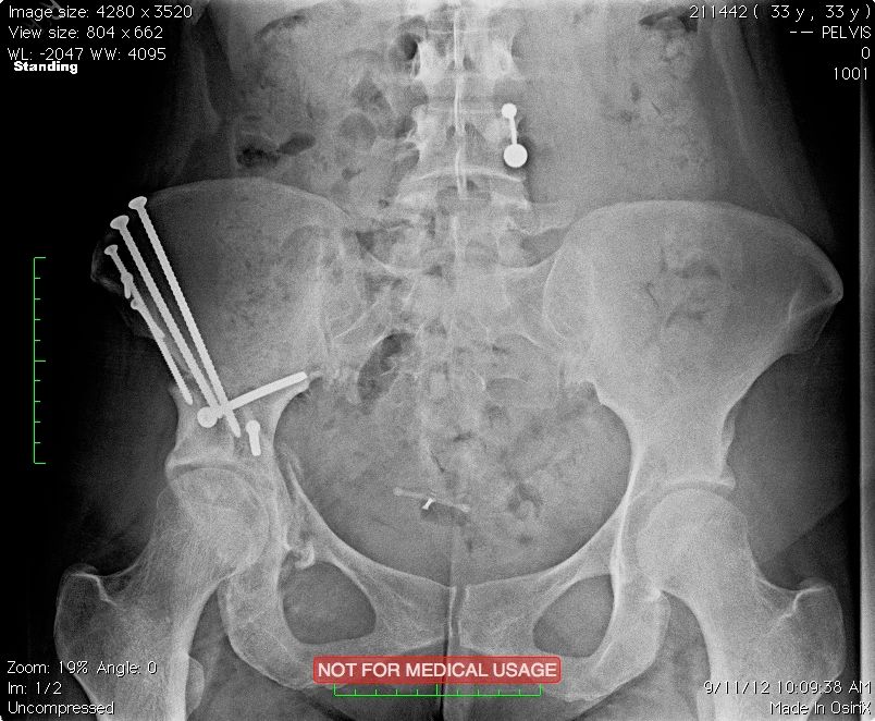
X-ray from September
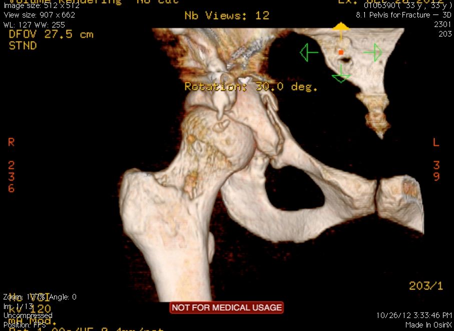
3D Image from CT showing Fracture of the Superior Pubic Ramis
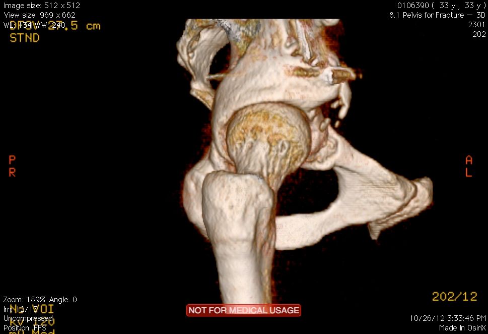
 3D Image from CT showing a small heterotrophic ossification HO (bone forming in the muscle)